Paper Chromatography

Paper chromatography has proved to be very successful in the analysis of chemical compound and lipid sample in particular.

Nature of the paper:

The paper commonly used consists of highly purified cellulose. Cellulose, a homopolysaccharide of glucose. Contains several thousand anhydro-glucose units-linked through oxygen atoms. The paper exhibits weak ion exchange and adsorptive properties. Modified forms of paper have been produced in which the paper has been impregnated with alumina, silica gel, and ion-exchange resin etc.

The chemical composition of whatmann filter paper no: 1 is: a-cellulose (98 to 99%), b-cellulose (0.3 to 1%), Pentosans (0.4 to 0.8%), Ash (0.07 to 0.1%) & ether soluble matter (0.015 to 0.1%).

Apparatus:

The apparatus required for paper chromatography arepaperchrom

1) Support for paper

2) Solvent trough

3) Airtight chamber

4) Whattmann filter paper number 1

5) Capillary tubes

6) Samples – Amino acids (or) Pigments

7) Solvents

8) Platinum loop

Paper development

There are two main techniques, which may be employed for the development of paper Chromatograms.

1) Ascending techniques

2) Descending techniques

3) Radial development

4) Two-dimensional chromatography

1) Ascending techniques:

clip_image002The filter paper is then dried and equilibrated by putting it into on airtight cylindrical jar, which contains an aqueous solution of a solvent. The most widely applicable solvent mixture is n-butanol: acetic acid: Water (4:1:5), which is abbreviated as BAW.

The sheet of paper is supported on a frame with the button edge in contact with a trough with solvent. The arrangement is contained in an airtight tank lined with paper saturated with the solvent to prove a constant atmosphere and separations are carried out in a constant temperature room. Thus, the solvent will ascend into the paper this process is, therefore, termed “Ascending Chromatography”

2) Descending techniques:

The end of the filter paper may be put into the solvent mixture contained in a narrow trough mounted near the top of the container. In this chromatography, the solvent will descend into the paper and this process is then termed “Descending Chromatography”.

clip_image005This method is convenient for compounds, which have similar Rf values since the solvent drips off the bottom of the paper, thus giving a wider separation.

3) Two dimensional chromatography (3D):

The mixture is separated then the first solvent, which should be volatile: then after drying, the paper is turned through 900 and separation is carried out in the second solvent.

After locating the migrated unknown sample along with standard known sample, a map is obtained and comparing their position with a map of known compounds can identify compounds.

Locating the compounds:

Strip is removed when the solvent has migrated over most of the available space. The distance to which the solvent has run is marked. In most cases, the completed Chromatogram is colorless with no indication of the presence of any compounds. Such a chromatogram is said as “Undeveloped” for locating the various compounds. The filter paper strip is first dried, then sprayed with 0.5% Ninhydrin in acetone and at least heated for a few minutes at 80 to 1000 C. the reaction occurs and the colored spots appear at the sites of the amino acids, such as Chromatogram is now called “Developed”.

In paper chromatography, the stationary cellulose phase is more polar than the mobile organic phase.

image

Identifying the compounds:

The ratio of the distance travelled by a component (i.e. amino acid) to that travelled by the solvent front, both measured from the marked point of the application of the mixture, is called the “Resolution front (Rf)” value for that component.

   Distance from origin run by the compound

Rf = -----------------------------------------------------------------------------

Distance from origin run by the solvent


chromatography_diagram

Detection:

The filter paper strip may be sprayed with ninhydrin and heated so that the colored spots indicating the location of amino acids may develop. The color densities of these spots may be measured with a recording transmittance (or) reflectance photometer device.

ninhydrin01

Ninhydrin test: ninhydrin

  • Amines (including α-amino acids) react with ninhydrin to give a coloured product.
  • It can be used qualitatively (e.g. for chromatographic visualisation) or quantitatively (e.g. for peptide sequencing).
  • The α-amino acids typically give a blue-purple product.
  • Proline, a secondary amine, gives a yellow-orange product.
  • The test is sensitive enough that ninhydrin can be used for the visualisation of fingerprints.

Applications:

By using this technique

1) To check the control of purity of pharmaceuticals,

2) To the detection of adulterants,

3) To detect the contaminants in foods and drinks,

4) To the study of ripening and fermentation,

5) To the detection of drugs and dopes in animals & humans

6) To the analysis of cosmetics

7) To the analysis of the reaction mixtures in biochemical labs.

THIN LAYER CHROMATOGRAPHY (TLC)

TLC may be either carried out by the adsorption principle (if the thin layer is prepared by an adsorbent such as “Keiselguhr” (or) “Alumina” (or) by the partition principle (if the layer is prepared by a substance such as “Silica gel” which hold water like the paper).

Chromatography _Paper2

Preparation of the layer:

The glass plate should be washed thoroughly & dried before layer application. The material to be used for layer preparation is a follows:

The selected material is usually mixed with water, it form thick suspension, known as ”Slurry”. This slurry is applied to a plate surface uniformly with 0.25mm thickness. To this layer mix the binder “Calcium sulphate” for better adhesion of the stationary phase. The plates are dried after application of the slurry. If adsorption chromatography is to be performed, the thin layer is activated by heating at 1100 C for several hours.

Compounds

Adsorbents

Solvent systems

Mono and Disaccharides

Kieselguhr.G

(Sod.acetate)

Kieselguhr.G

(Sod.Phosphate, pH-5.0)

Ethylacetate/Propanol

(65/35)

Butanol/Acetone/Phosphate buffer (pH-5.0)

(40-50-10)

Amino acids

Silica Gel.G

96% ethanol/water

(70/30)

Plant pigments

Kieselguhr.G

Petroleum ether/Propanol

(99/1)

Procedure:spotting

Chromatographic plates (20X20cm) of 200m thicknesses are prepared by using a suspension of 30 grams of silica gel G in 63ml of 0.1M Na2CO3 solutions by shaking vigorously for 90 seconds. The silica gel slurry is applied on to the glass plate in the form of a uniform layer. These plated are activated at 1100C for 30 minutes immediately prior to use.

Then the samples (5 to 100mL) are applied on the silica plate in the form of small drops at regular intervals. In this plate these samples are applied as a spot of less than 5 minutes diameter on the lower right corner of the plates under a stream of warm air.Thin Layer CHROMATOGRAPHY Plate2

This plate is introduced into the saturated standard Brinkman developing chamber with the vapor of the solvent mixture with Chloroform: Methanol: Acetic acid: Water (250:74:19:3 v/v) to dip 4.5 cm of its bottom.

tlc (1)

When solvent migrates about 15cm, plates are dried in air for 15 minutes and develop in the second dimension (900 rotation clockwise) with CHCl3: CH3OH: 7M NH4OH (230: 90: 50 v/v). The solvent front is again allowed to stand (or) more about 15cm. Then the plate is dried in air for 5 minutes and exposed to iodine vapor (or) UV light the sample molecules can be visualized. When a permanent record of developed plates is desired, plates are sprayed lightly with 10N H2SO4 and then heated at 1100 for 15 minutes.TLC

By calculating the Rf values are can easily identified the molecules present in the mixture.

Detection: Several detection methods are available. They are,

1) Spraying the plate with 25 to 50% H2SO4 in ethanol and heating. This results in charring of most of the compounds, which show up as Brown spots.

2) Iodine vapours are used extensively as a universal reagent for organic compounds. This iodine spot disappears rapidly but can be made more permanent by spraying with 0.5% benzidine solution in absolute ethanol.

Applications:

1) The constituents of the mixture of amino acids, and the constituents of natural lipids and phospholipids are separated and estimated in a short time.

2) Enzymes, nucleic acids, pigments, sugars can also be separated by using this technique.

3) TLC has often been used to identify drugs, contaminants & Adulterants.

Advanced TLC:

TLC can be automated using forced solvent flow, running the plate in an vacuum-capable chamber to dry the plate, and recording the finished chromatogram by absorption or fluorescence spectroscopy with a light source. The ability to program the solvent delivery makes it convenient to do multiple developments in which the solvent flows for a short period of time, the TLC plate is dried, and the process is repeated. This method refocuses the spots to acheive higher resolution than in a single run. See for example: Poole, C. F.; Poole, S. K. "Instrumental Thin-Layer Chromatography," Anal. Chem. 1994, 66, 27A.

Two-dimensional TLC uses the TLC method twice to separate spots that are unresolved by only one solvent. After running a sample in one solvent, the TLC plate is removed, dried, rotated 90o, and run in another solvent. Any of the spots from the first run that contain mixtures can now be separated. The finished chromatogram is a two-dimensional array of spots.

Gel Filtration

This is also known as “Molecular exclusion chromatography” (or) “Molecular sieve chromatography”, “Size exclusion chromatography” and “Permeation chromatography

Principle:

In exclusion chromatography the separation of molecules is based up on the size and shape. The stationary phase is porous bead material and the mobile phase is the solvent system.

The large molecules cannot enter the pores of the beads so they are excluded out and come down rapidly. Small sized molecules enter the pores of the beads so that their speed is retarded and comedown slowly. The degree of retardation of a molecule is proportional to the time it spends inside the gel pores, which is a function of the molecule’s size and the pore diameter.

gel filtration

Mathematical relationships about solute behaviour on molecular sieve gels:

1) “The distribution of a solute particle between the inner and outer solvent (solvent within and gel filtrationoutside of the gel bead) is defined as “Distribution coefficient” (Kd).

Kd= 0 –> Solute molecule is large & excluded out completely

Kd= 1 –> Solute molecule is small, retards it in inner solvents

2) The volume of outer solvent, i.e., the solvent surrounding the gel beads is indicated as Vo. The technical term for this is “Void volume”.

The volume of solvent inside gel bead – Inner solvent = Vi

The distribution coefficient = Kd

The effluent volume = Ve

Thus

clip_image001

The volume of inner solvent, Vi, can be calculated if the dry weight of gel employed and the “Water regain value” of the particular gel are known. Thus,

clip_image003clip_image002

  • For two different substances possessing different molecules weights and therefore, different distribution coefficients (Kd1 & Kd2) the difference in their effluent volumes, Vs is given by


Vs =Ve1-Ve2 = (V0+Kd1.Vi)-(Vo+Kd2.Vi)clip_image005


Types of gels:

Characteristics of gels:

1) The gel material should be chemically inert.

2) It should preferably contain ravishingly small number of ionic groups.

3) Gel material should provide a wide choice of pore and particle sizes.

4) The gel should have uniform particle & pore sizes.

5) The gel matrix should have high mechanical rigidity.

There are FIVE principle types of media:

a) Sephadex

b) Agarose

c)Polyacrylamide (Bio-gel.P)

d) Styragel e) Porous glass & silica granules

a) Sephadex:

1. It is most popular gel for proteins & most of the biomolecules separation.

2. When”Leuconosto mesenteroids” indulge in sucrose fermentation, large polymers of glucose are the results. These polymers are known as “Dextrans” are used to prepare sephadex.

3. It is cross-linked polymer.

4. Sephadex gels are insoluble in water, and are stable in bases, weak acids and mild reducing and oxidizing agents.

5. Sephadex gels are insoluble in water, and are stable in bases, weak acids and mild reducing and oxidizing agents.

6. Sphadex, which cannot be used to separate biopolymers larger than 300,000 Daltons.

7. Some identified gels serial number gels are G-25; G-50; G-75; G-100; G-200

b) Agarose:

1. Agarose gels are produced from AGAR.

2. They are linear polysaccharides alternating residues of D-Galactose and 3,6-anhydro-L-galactose units.

3. These gels are hydrophilic and are almost completely free of charged groups.

4. Agarose gels, due to their greater porosity, it may be used to separate molecules and participates up to a molecular weight of several million Daltons.

5. The gels are used in the study of viruses, nucleic acids and polysaccharides.

6. Some commonly used agarose gels are

--> Sepharose 2B

--> Sepharose 4B

--> Sepharose 6B

c) Polyacrylamide:

1. This is very popular medium is produced by polymerizing acrylamide into bead form.

2. polyacrylamide gels can be used to separate molecules of up to 300,000 daltons.

3. This gel is insoluble in water and common organic solvents may be used in the pH range of 2 to 11.

4. Some common gels are,

Bio-gel P 10,    Bio-gel P60,   Bio-gel P100,

Bio-gel P200,  Bio-gel P300

d) Styragel:

1. For completely non-aqueous separations, a gel that will swell in an organic solvent is required. Styragel provides this option.

2. It is a rigid cross-linked polystyrene gel.

3. The gel structure is unaffected by temperatures as high as 1500 C

4. the gel can be used with such solvent as tetrahydrofuran, cresol, dimethyl sulfoxide, chloroform, carbon tetrachloride and others.

e) Controlled pore glass beads:

1. These fine glass spheres are manufactured from borosilicate glass to contain large number of pores within a very narrow size distribution.

2. The glass spheres have a molecular exclusion limit ranging from 3000 to 9 million Daltons.

Procedure:

This is mainly carried out in the columns. The column is filled with beads, these beads contain pores. The beads are made up of gels. The gels are made up of dextrans (Sephadex), Agarose (Biogel-A) and Polyacrylamide (Biogel-P).

The sample is discovered in buffer and allowed to flow through the column. Large molecules can not enter the pores of the beads, they are excluded out and reach down fastly. The small sized molecules enter into the pores of the beads; their speed is retarded and reaches down slowly. These particles are analyzed by spectroscopy.

gel filtration

Applications:

1) The main application of gel-filtration is the purification of molecules, viruses, nucleic acids, hormones, enzymes, proteins, and antibodies and can be separated and purified by this technique.

2) It is also used for the separation of vitamins, steroids, neuropeptides and drugs.

3) Separations are achieved very quickly by this technique.

4) The molecular weight of the molecule can also be determined by this technique.

5) Protein receptor binding can be understood by this technique.

6) This method is especially useful for the separation of 4S and 5S tRNA.

7) It is also the most satisfactory method for separating DNA (from bacteria, usually Gram positive) from the invariable contaminants, the “Teichoic acid”.

CHROMATOGRAPHY

Chromatography is a technique used for separating or identifying the components in a mixture. It is a powerful method in industry, where it is used on a large scale to separate and purify the intermediates and products in various syntheses. There are several different types of chromatography : 

  • Paper chromatography
  • Thin layer chromatography (TLC)
  • Gas chromatography (GC)
  • Liquid chromatography (LC)
  • Ion exchange chromatography
  • Affinity chromatography

Basic principle

All chromatographic methods require one static part called “the stationary phase” and one moving part “the mobile phase”. The techniques rely on one of the following phenomena: adsorption; partition; ion exchange; or molecular exclusion.

Adsorption

Adsorption chromatography was developed first. It has a solid stationary phase and a liquid or gaseous mobile phase. Each solute has its own equilibrium between adsorption onto the surface of the solid and solubility in the solvent, the least soluble or best adsorbed ones travel more slowly. The result is a separation into bands containing different solutes. Liquid chromatography using a column containing silica gel or alumina is an example of adsorption chromatography.

Eluent:

The solvent that is put into a column is called the eluent.

Eluate:

The liquid that flows out of the end of the column is called the eluate.

Partition

In partition chromatography the stationary phase is a non-volatile liquid which is held as a thin layer on the surface of an inert solid. The mixture to be separated is carried by a gas or a liquid as the mobile phase. The solutes distribute themselves between the moving and the stationary phases, with the more soluble component in the mobile phase reaching the end of the chromatography column first. Paper chromatography is an example of partition chromatography.

Types of chromatography

1. Paper chromatography

In paper chromatography, the sample mixture is applied to a piece of filter paper, the edge of the paper is immersed in a solvent, and the solvent moves up the paper by capillary action. Components of the mixture are carried along with the solvent up the paper to varying degrees, depending on the compound's preference to be adsorbed onto the paper versus being carried along with the solvent. The paper is composed of cellulose to which polar water molecules are adsorbed, while the solvent is less polar, usually consisting of a mixture of water and an organic liquid. The paper is called the stationary phase while the solvent is referred to as the mobile phase. In order to obtain a measure of the extent of movement of a component in a paper chromatography experiment, we can calculate an "Rf value" for each separated component in the developed chromatogram. An Rf value is a number that is defined as: distance traveled by component from application point

Paper chromatography


2. Thin layer chromatography (TLC)

Thin layer chromatography (TLC) is a method for identifying substances and testing the purity of compounds. Separations in TLC involve distributing a mixture of two or more substances between a stationary phase and a mobile phase. The stationary phase is a thin layer of adsorbent (usually silica gel or alumina) coated on a plate. The mobile phase is a developing liquid which travels up the stationary phase, carrying the samples with it. Components of the samples will separate according to how strongly they adsorb on the stationary phase versus how readily they dissolve in the mobile phase.TLC chamber

3. Gas Chromatography:

Gas chromatography makes use of a pressurized gas cylinder and a carrier gas, such as helium, to carry the solute through the column. The most common detectors used in this type of chromatography are thermal conductivity and flame ionization detectors. There are three types of gas chromatography that will be discussed here: gas adsorption, gas-liquid and capillary gas chromatography. Gas adsorption chromatography involves a packed bed comprised of an adsorbent used as the stationary phase. Common adsorbents are zeolite, silica gel and activated alumina. This method is commonly used to separate mixtures of gases.

Gas Chromatography

4. Liquid Chromatography

There are a variety of types of liquid chromatography. There is liquid adsorption chromatography in which an adsorbent is used. This method is used in large-scale applications since adsorbents are relatively inexpensive. There is also liquid- liquid chromatography which is analogous to gas-liquid chromatography. The three types that will be considered here fall under the category of modern liquid chromatography. They are reverse phase, high performance and size exclusion liquid chromatography, along with supercritical fluid chromatography. Reverse phase chromatography is a powerful analytical tool and involves a hydrophobic, low polarity stationary phase which is chemically bonded to an inert solid such as silica. The separation is essentially an extraction operation and is useful for separating non-volatile components.

Liquid Chromatography

High performance liquid chromatography (HPLC) is similar to reverse phase, only in this method, the process is conducted at a high velocity and pressure drop. The column is shorter and has a small diameter, but it is equivalent to possessing a large number of equilibrium stages. Size exclusion chromatography, also known as gel permeation or filtration chromatography does not involve any adsorption and is extremely fast. The packing is a porous gel, and is capable of separating large molecules from smaller ones. The larger molecules elute first since they cannot penetrate the pores. This method is common in protein separation and purification.

Supercritical fluid chromatography is a relatively new analytical tool. In this method, the carrier is a supercritical fluid, such as carbon dioxide mixed with a modifier. Compared to liquids, supercritical fluids have solubilities and densities have as large, and they have diffusivities and viscosities quite a bit larger. This type of chromatography has not yet been implemented on a large scale.

5. Ion Exchange Chromatography

ion-exchange chromatography

Ion exchange chromatography is commonly used in the purification of biological materials. There are two types of exchange: cation exchange in which the stationary phase carries a negative charge, and anion exchange in which the stationary phase carries a positive charge. Charged molecules in the liquid phase pass through the column until a binding site in the stationary phase appears. The molecule will not elute from the column until a solution of varying pH or ionic strength is passed through it. Separation by this method is highly selective. Since the resins are fairly inexpensive and high capacities can be used, this method of separation is applied early in the overall process.

6. Affinity Chromatography

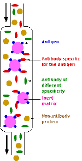
Affinity chromatography involves the use of packing which has been chemically modified by attaching a compound with a specific affinity for the desired molecules, primarily biological compounds. The packing material used, called the affinity matrix, must be inert and easily modified. Agarose is the most common substance used, in spite of its cost. The ligands, or "affinity tails", that are inserted into the matrix can be genetically engineered to possess a specific affinity. In a process similar to ion exchange chromatography, the desired molecules adsorb to the ligands on the matrix until a solution of high salt concentration is passed through the column. This causes desorption of the molecules from the ligands, and they elute from the column. Fouling of the matrix can occur when a large number of impurities are present, therefore, this type of chromatography is usually implemented late in the process.


Affinity chromatography

ION-EXCHANGE CHROMATOGRAPHY

W.cohn first developed this procedure. The reversible exchange of ions in solution with ions electrostatically bound to some sort of insoluble support medium.

Principle:
Exchange of ions is the basic principle in this type of Chromatography. In this process two types of exchangers i.e., cationic and anionic exchangers can be used.
Cationic exchangers possess negatively charged group, and these will attract positively charged cations. These exchangers are also called “Acidic ion exchange materials”, because their negative charges result from the ionization of acidic group.

Ion Exchange Chromatography
Anionic exchangers have positively charged groups that will attract negatively charged anions. These are also called “Basic ion exchange” materials.

Ionic Exchangers
 
 
Types of ion exchange resins:

  • Two main groups of materials are used to prepare ion exchange resins: Polystyrene and Cellulose.
  • Resins made from both of these materials differ in their flow properties, ion accessibility and chemicals and mechanical stability.
  • Polystyrene resins are proposed by polymerization reaction of styrene and divinyl benzene.
  • A higher concentration of divinyl benzene produces higher cross linkages.
  • 5. Polystyrene resins are very useful for separating small molecular weight compounds.
  • Increasing the cross linkage increases the rigidity, reduces swelling, reduces porosity & reduces the solubility of the polymeric structure.
  • sulfonic acids are strong acids with good proton dissociation ability. By sulfonation process, acidic functional groups are easily attached to nearly every aromatic nucleaus.
  • Resins substituted with sulfonic acid groups are strong cationic exchangers.
  • To prepare weekly acidic exchanger, carbohydrate groups can be attached to the aromatic rings instead of sulfonic acid group.
  • If basic functional groups are introduced, the resin can exchange anions rather than cations. Strong anion exchangers are prepared with a tertiary amine, yielding a strongly basic quaternary ammonium group. Weak anionic exchangers can be prepared with secondary amines, yielding a weakly basic tertiary amine.


Cellulose resins have much greater permeability to macromolecular polyelectrolytes and possess a much lower charge density as compared to polystyrene exchangers.
  • Carboxymethyl cellulose (CM-cellulose) – Cationic exchanger
  • DEAE cellulose - Anionic exchanger
Preparation of the exchange medium:
There are three steps are of absolute importance:
1) Swelling of medium: (Pre-cycling):

Swelling makes the functional groups to be exposed for ion exchange.
    • Swellimg of anion exchangers is usually carried out by treating it. first with an acid (0.5N HCl) and then with base (0.5N NaOH).
    • Exactly the reverse is the case with cationic exchangers. The matrix can be treated with EDTA for impurity eliminations.

2) Removal of very small particles:
These fines will decrease flow rate and unsatisfactory reaction. To remove fines, the exchanger is repeatedly suspended in a large volume of water and after the larger polymers have settle down, the slow sedimenting materials decanted.

3) Equilibration with counter ions:
This is accomplished by washing the exchanger with different reagents depending upon the desired counterion to be introduced.
    • NaOH –> counter ion to be introduced is “Na+
    • HCl –> counter ion to be introduced is “H+
    • NaNO3 –> counter ion to be introduced is “NO3
Choice of Buffers:
  • Anionic exchange Chromatography should be carried out with cationic buffers.
  • Cationic exchange Chromatography should be carried out with anionic buffers.
  • The pK of the buffer should be as near as possible to the pH at which the system is buffered. This results in high buffer capacity, which can with stand the local changes of pH in the column easily.

    Buffers


    PH range
    Ammonium acetate 4 to 6
    Ammonium formate 3 to 5
    Pyridinium formate 3 to 6
    Pyridinium acetate 4 to 6
    Ammonium carbonate 8 to 10

Practical procedure:
Ion exchange separations are carried out mainly in columns packed with an ion-exchanger. These ionic exchangers are commercially available. They are made up of styrene and divinyl benzene.

DEAE-cellulose is an anionic exchanger, CM-cellulose is a cationic exchanger. The choice of the exchanger depends upon the charge of particle to be separated. To separate anions “Anionic exchanger” is used, to separate cations “Cationic exchanger” is used.
First the column is filled with ion exchanger then the sample is applied followed by the buffer. The tris-buffer, pyridine buffer, acetate buffer, citrate and phosphate buffers are widely used. The particles which have high affinity for ion exchanger will come down the column along with buffers. In next step using corresponding buffer separates the tightly bound particles. Then these particles are analyzed spectroscopically.
Ionic Exchange chromatography
Applications:
1. It is extremely used in the analysis of amino acids. The amino acid “Autoanalyzer” is based on in exchange principle.
2. To determine the base composition of nucleic acids. Chargaff used this technique for established the equivalence of Adenine and Thymine; Guanine and Cytosine.
3. This is most effective method for water purification. Complete deionization of water (or) a non-electrolyte solution is performed by exchanging solute cations for hydrogen ions and solute anions for hydroxyl ions. This is usually achieved by method is used for softening of drinking water.
4. Proteins are also successfully separated by this technique.
5. It is also used for the separation of many vitamins, other biological amines, and organic acids and bases.


AFFINITY CHROMATOGRAPHY

It is mainly based on the biological affinity (or) biological specificity. This technique mainly requires previous knowledge of the molecule to be separated a specific ligand will only attach with a specific molecule.

clip_image001

The materials to be isolated are capable of binding reversibly to a specific ligand i.e., attached to an insoluble matrix.

affi

Why Use Affinity Chromatography?

Affinity chromatography offers high selectivity, resolution, and capacity in most protein purification schemes. It is the only technique that has the advantage of utilizing a protein's biological structure or function for purification. As a result, purifications that would otherwise be time consuming and complicated, can often be easily achieved with affinity chromatography.


Supporting matrix:

1) Characteristics of Matrix:

  • The matrix should be inert to other molecules to minimize non-specific adsorption.
  • It should possess good flow properties.
  • It should be chemically and mechanically stable at varying pH, ionic strength and denaturating conditions employed for binding and elution.
  • It should contain large numbers of suitable chemical groups for ligand attachment.
  • It should be highly porous a large surface area for attachment of the ligand and allows interaction of the desired macromolecule with the immobilized ligand.

2) Types:

The particles, which are uniform, spherical and rigid, are used. The most commonly used ones are

a) Agarose                  b) Polyacrylamide                  c) Controlled glass beads

a) Agarose:

The agarose beads have most desired features as mentioned above. But it has some advantage when use the denaturant solution for elution, which have a susceptibility to contraction.

b) Polyacrylamide:

The polyacrylamide bead lacks porosity. This undesirable trait is heightened even further when they are substituted by ligands.

c) Controlled porosity glass beads:

This bead provides mechanical rigidity and chemical inertness in addition to providing very good flow rates. High degree of nonspecific protein adsorption is the most serious drawback to these beads, which avoid to some extent by treatment with “Hexamethyldisilazane”.

3) Ligand selection:

The selection of ligand should have two most important requirements:

    • Ligand interaction should be less with desired macromolecules.
    • The ligand should possess functional groups that can be modified to form covalent linkage with the supporting matrix.

4) Ligand attachment:

Covalent coupling of the ligand to the supporting matrix involve the following steps:

    • Activation of the matrix functional groups
    • Covalent attachment of the ligand to the activated functional groups.

i) Activation of the matrix functional groups:

The most common method of activation of polysaccharide supports (agarose) involves treatment with “CNBr” at alkaline pH (pH=11.0). Usually 300 mg of powdered cyanogens bromide used per ml of packed gel gives the maximum substitution. The reaction is exothermic and maintains the temperature constant at 200C at all times. To maintain temperature the pH at 11, the mixture is continuously stirred and an electrode dipped into it at all times. The pH is maintained by the addition of 2M NaOH. The activated suspension is now washed with about 20 times the gel volume with a buffer (buffers –Tris, Ammonium acetate, Glycine) at a pH of 9.5 to 10. Usually just 10 to 15 minutes are required for the reaction to be completed. Sodium bicarbonate and borate buffers are the usual choice.

ii) Covalent attachment of the ligand to the activated functional groups:

Coupling of amino – containing ligand to CNBr activated support is normally carried out by suspending the support and the ligand in a basic buffer solution at pH (0.25 M NaHCO3, pH-9.0). The suspension stirred overnight in a cold room. During this time the ligand is covalently attached to the support medium.

After the reaction is over, the matrix should wash with 0.1 M solution of pH 9.0 glycine buffer, the solution destructs the any extra-activated groups.

The number of ligand group bound is usually expressed in terms of capacity per ml of packed matrix rather than in terms of its dry weight.

 Some group specific ligands

image

5) The ARM:

To avoid the encounter steric repulsion between ligand and activated groups of matrix with macromolecule, which is used to introduce a spacer between the activated groups of the support and the ligand. This space is known as “ARM”. The ligand projects out the macromolecule to prevent repulsion.

E.g.:

1)Hexamethylene, 3,3’-diamino propylamine

2) 1,6-diamino hexane

3) 6-amino-hexanoic acid

4) 1,4-bis-(2,3-epoxypropoxy) butane

These spacer arms have two different functional groups; one to react with the functional groups of the matrix and the other is to react with ligand.

In organo synthetic procedures “Succinic anhydride” and a “Water soluble carbodiimide” are using to attaches the ligand.


Practical Procedure:

It is also carried out in the column. In this matrix and ligands are used. Prior to use, the gel (or) matrix must be converted to the swollen form, done by allowing a known weight of the gel to swell either in water (or) in a weak salt solution. The greater the porosity, the more will be the time required to reach equilibrium.chromt11_img1

Agarose reacts with cyanogens-Bromide and forms activated complex. Then this activated complex reacts with epoxide and forms agarose cyanogens bromide-epoxide complex. Epoxide is a spacer arm attaches the matrix with ligands.

The agarose-CNBr-epoxide complex is filled in column. Then the specific ligand is added. Now the ligand react with the spacer arm and attaches to it. Then the sample is applied to the column. The specific ligand attaches with specific molecules. The remaining material will comedown the column, the attached molecule can be obtained by suitable buffer. The buffer supplement on the gel bed, it encourages adsorption of the desired molecule the buffer chosen must be supplemented with any cofactors (e.g.: Metal ions) required for “Ligand-Macromolecule interaction”. The buffer should also possess a high ionic strength so as to minimize non-specific polyelectrolyte adsorption onto charged groups in the ligand.

Applications:

    1. The technique has been used to purify a large variety of macromolecule such as enzymes, Immunoglobulins, membrane receptors, Nucleic acids and even polysaccharides.
    2. By using affinity chromatography, Whole cells have been purify include fat cells, T and B-lymphocytes, Spleen cells, Lymph node cells, Oocytes and chick embryo neural cells.
    3. Metal chelate affinity chromatography is the logical extension technique. Same molecular weight protein can be separated by this technique by using the metal ion containing matrix by chelation, because of their difference in their metal binding ability with proteins.
    4. By using the “Magnetic gel beads affinity chromatography”, immunoglobulin negative thymocytes and neuroblastoma cells have been purified by this method. The magnetic gel beads, usually polyacrylamide (or) agarose have a core made up of Fe3 O4 (Magnetite) and are chemically coupled to a protein ligand.
    5. Immobilized enzymes (Solid-state enzymes) are also isolated and purified by this method.
    6. mRNA can be isolated by this technique.
    7. Native proteins can be separated from denatured proteins by this technique.
    8. DNA & RNA can be separated from each other
    9. Papain and Urease can be separated by this technique.